Here is the translation of the text you provided:
Inside a Bunker Underneath the Jerusalem Hospital Lies a Particle Accelerator, Cyclotron by Name
Dror Feuer entered the holy of holies and tried to understand how they produce such innovative radioactive materials here, which, after being injected into cancer patients, will act in the body like guided missiles on their way to locate and eliminate targets.
Special for G
The soundtrack four floors below ground, in the basement of the Nuclear Medicine Department at Hadassah Ein Kerem Hospital, is one I’ve heard during thousands of hours of watching science fiction series and movies. It’s like being on the bridge of the Starship Enterprise. The noises from the world above don’t reach here. Here, you only hear mechanical beeps and digital rings. Beep-beep, cling-cling, and of course, woo-woo and ee-ee and wrrrrrr, and above all, there’s a thin layer of continuous hissing.
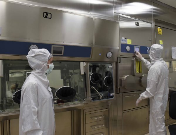
Here, between 2.5-meter-thick walls with cooling and air conditioning systems isolating it from the rest of the hospital, lie two particle accelerators, cyclotrons by name. One is old, about twenty years old, and the other is newer, about seven years old. They look the same. Each one resides in its own bunker, with its own control and monitoring room, cooling, maintenance, and electronics rooms. On the way there, we pass through and I derive great pleasure from the charming contrast between the high-tech and the delightful chaos in the place. Piles of papers lie everywhere, boxes, instruments, bottles, and whatnot. A small coffee corner leads to the maintenance room full of saws, hammers, screwdrivers, and a variety of tools I didn’t expect to see in a particle accelerator. Very Israeli. The control rooms are full of clocks and measuring instruments, some of which are going forward, and some backward.
We enter the holy of holies itself. The accelerator occupies most of a small room, with thousands of pipes in all colors and diameters coming in and out, looking like the forbidden yet passionate lovechild of a spaceship and a pressure cooker. A little over 2 million dollars each. It looks a little like a living creature. I can’t decide if I expected more or less, in any case, I’m satisfied. Everywhere I touch, I’m immediately told: “Don’t touch!” and then they walk through with the radiation meter, which beeps happily. Everything’s fine, they reassure me, nothing dangerous. So every time they’re not looking, I touch.
Inside the accelerator, there is a vacuum, like in space, the result of four diffusion pumps that export a pressure of 10 to the minus 7 atmospheres. “Explain to me what’s happening here,” I ask Professor Eyal Meshani, head of the cyclotron unit. “To make a nuclear reaction,” he responds, “we take charged particles, accelerate them in an electric and magnetic field, and then bombard them with other stable materials.”
Clear. Now explain it to me slower.
“First, you create a charged particle. In our case, we take hydrogen gas, put it inside, and pass it through a high electric field. It breaks it down and creates plasma. Plasma is chaos. From it, I extract, using another electric field, another particle that, because it has mass and electric charge, starts moving in a circular motion inside the electric and magnetic field. With each rotation, it increases in speed and energy until it reaches an energy of 18 mega-electron volts, and then I shoot it out of the field into a target, where it’s bombarded with high-energy protons, creating a new element called F19. That’s the radioactivity.
“Think that inside this insane vacuum, there are no collisions, and I can focus a beam to millimeters,” Meshani continues. “The whole process is cooled with helium to minus 180 degrees. What comes out, a radioactive solution, I send through thin tubes to the lab, where I separate the radioactivity from the water and charge it onto a smart molecule.”
“It’s True for Many Diseases”
Meshani, 54, probably sees in my eyes that while my body is here, my mind is still catching up. “Let’s say you have a military hangar in Syria,” he demonstrates. “You send a plane that photographs it from above, and now you know everything about the configuration of the place, its location, the setup, the fences, etc. But you don’t know anything about what’s inside, whether it’s tanks, planes, or missiles. That’s actually anatomical imaging, like X-ray, CT, ultrasound, and also MRI. But that’s not enough, right?”
Of course not.
“The world is moving towards personalized and targeted medicine. This requires much more complex and intelligent solutions, but for that, we need information. How do we get it? There’s an invasive way—cutting, doing a biopsy, taking samples, doing pathology. It’s great, but there are problems with it—it takes time, you need to take samples from different places, and it’s painful. There’s another way to get information—molecular imaging.
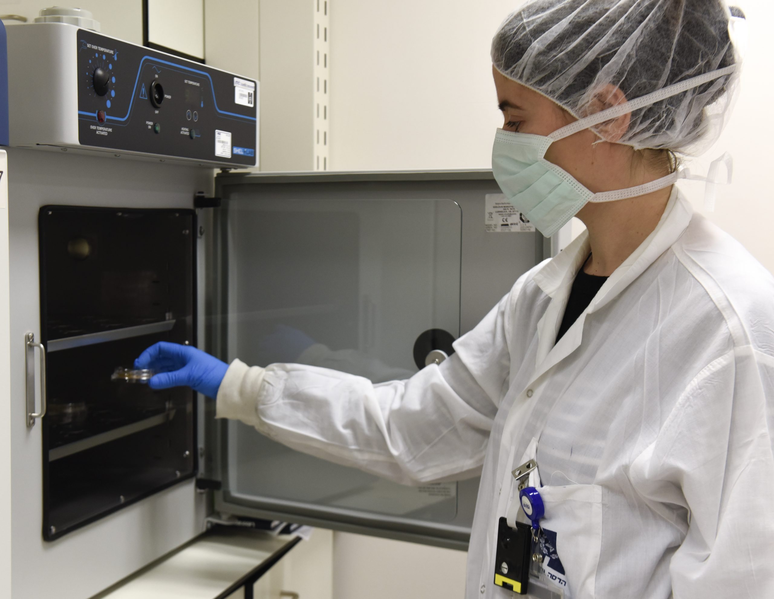
“To go back to the analogy, instead of sending a plane, I send Sayeret Matkal (Israeli elite unit), who enter and report exactly what’s going on inside. That’s molecular imaging, which allows us to see elements at the cellular level. How? You inject a radioactive material, which is a smart material. A guided missile. You put the material into the blood circulation, and it’s attracted to certain elements of the tumor. These materials are what we produce here, in the accelerator. I’m essentially inserting a spy program into tissue in the human body that constantly sends me signals. The smart material knows where to go, and the radioactive isotope knows what to do there.”
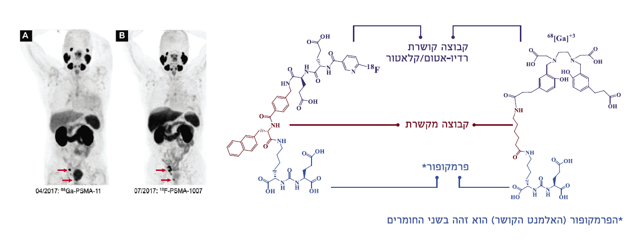
In such an imaging, PET-CT, unlike anatomical imaging, you can see very precisely, for example, if there are certain lung tumors that only 20% of the patients have. Then, instead of bombarding the whole body with everything you have, you can take smart materials and load them with a radioactive isotope that you inject into the blood. “The isotope sends us signals, and we track it when it accumulates in the tumor area and then know if the element we want to bombard is there or not,” says Meshani. “And this is true for many diseases. For example, understanding processes in the brain, like in the case of Alzheimer’s, where beta-amyloid plaques accumulate there, and there are materials that can find these plaques long before the symptoms start. This is excellent for early diagnosis, even though there is still no cure, and we produce these materials here.”
If you’ve already reached the tumor, why just send signals and not blow it up?
“If there are neuroendocrine tumors (see glossary to the left), we treat them with a material that knows how to reach them and kill them. That’s the direction, but it’s important to emphasize, this doesn’t replace biopsies or other treatments.”
And in the future?
“Like everything, there will be a combination of tools. It’s one of the tools in modern medicine, to guide more effective drug treatment and to track it better. Another advantage of this imaging is that it’s much faster than CT or ultrasound. If you give a biological substance in a CT to kill a tumor, you can only see the result when the mass disappears, and that can take four months. Here you can see it immediately. But it’s complicated, complicated, expensive, and not simple.”
What about dangerous?
“We give the material in very small quantities. In general, these materials have no mass, only electric charge. So, there’s no toxicity problem. The radiation is very specific, and we know how to read it. These are very unique materials, many of them don’t live long. They have a half-life. Some live for two minutes or less. The longest half-life of our materials here is 110 minutes. Such a half-life means you can’t import it or store it—you need daily production.”
The Difference Between a Reactor and a Cyclotron
We stand before the accelerator, which is currently working at 515 milli-amps, the pressure inside it reaches 31 bars, and the protons are bombarded at 75 micro-amps. “Today, we produced 5.5 curies, which is a lot,” says Meshani. It produces radioactive materials—mostly those that contain glucose, which is consumed in high percentages in the brain, bladder, and tumors. The material is injected into the body and tracked using the imaging device until it reaches the cancer cells, allowing for diagnosis and treatment.
The cyclotron uses both an electric and magnetic field to produce nuclear materials for research and medical uses, specifically for PET imaging. It was invented in the 1930s and consists of two half-cylinders in the shape of a “D,” between which is an electric field that accelerates charged particles that pass through in a circular motion (thanks to a magnetic field).
The accelerator keeps doing its thing, but what gives me pleasure is discovering that it’s all related to radiation—nuclear medicine from the future, but in the end, it’s still connected to the trash can. In the corner of the room, two trash bins—one for regular waste and one for radioactive waste. I hope they don’t get confused. “Before opening the cyclotron,” a large sign reads, “Don’t forget to turn off the air conditioner.”
Why are we so deep underground and between such thick walls? Could this thing explode?
“No. The beauty of the cyclotron, and the difference between a nuclear reactor and a particle accelerator, is that when you turn it off, there’s nothing. In Canada, they have accelerators inside shopping malls.”
So, how does it work on a daily basis, and how do you know what to produce? Do the hospitals call and say, ‘


